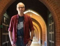
Christine remembers the moment when she first saw evidence that proved her ideas were right.
“Oh my goodness, I thought, we’ve just opened up a whole lot of new biology here. It’s like the oyster diver. Finally, you find the oyster with the pearl in it.”
She tells us about her journey of discovery.
The complexity and specificity of the developing brain is extraordinary. In the first three months of human gestation something amazing happens. Well over a million cells in the retina each send out a long thread-like nerve fibre, called an axon, that travels for up to three weeks through the optic chiasm to the back of the brain. There they each find other specific neurons in the brain with which they make synaptic connections. I studied this process mostly in frog embryos, where this entire process takes just 18 hours.
My fascination for living things started from a very young age. My brother and I would wake at dawn to explore the streams and fields where we lived in Northumberland. I could see the beauty of a caddisfly larvae and wondered how it came to be that way.
I remember vividly the moment I knew I wanted to understand nature at a deeper level. I was in my final year at Sussex University and everything I was being taught in molecular biology, genetics, developmental biology and neurobiology suddenly came together. I wrote an essay on how the visual system develops and I had questions, questions, questions. My love for understanding how the brain wires up had begun.
Different academics were coming up with very different theories for how the extremely specific connections between the eye and brain develop. But actually the techniques to answer the question just hadn’t yet been invented.
I was cycling along Oxford Street in London when the idea hit me of how we could do it. I was a PhD student at King’s College London with Professor John Scholes. We’d already discovered that we could take tiny pieces of embryonic frog eye tissue, incubate them in a radioactive nucleotide to ‘label’ the DNA, put them back and see only the cells that had been exposed and follow how they rearranged themselves during embryonic development. I realised that instead of labelling the DNA, I could label the growing axons with a radioactive amino acid.
It worked beautifully. Suddenly I was able to see embryonic axons growing from all these different quadrants of the eye. For the first time, it became clear that axons grow directly to their far-away targets rather than taking random walks and arriving there by trial and error.
It didn’t feel transformative at the time, but it turned out to be. Nature published my PhD work in two papers, which was amazing. I moved to the University of California San Diego to work with Bill Harris and spent the 1990s trying to identify the signposts in the embryonic brain that direct axons on their journey.


